


Digital twins
We transform complex diagnoses into surgical certainties through high-fidelity digital models
Surgical Planning Solutions
The evolution of diagnostic imaging
Digital twins allow for total immersion, where medical data is transformed into a tool for real-time prediction and surgical planning.
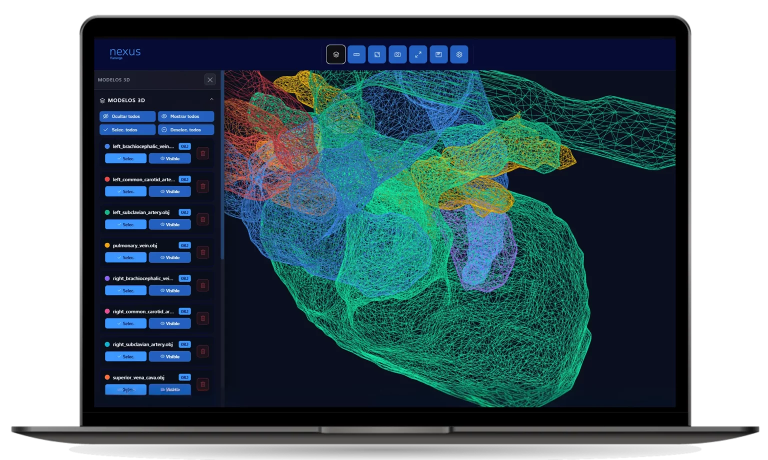
Advanced surgical planning software
Nexus is surgical planning software designed to eliminate surgical errors, provide faster diagnoses, and enable surgeons to perform complex procedures.
This tool provides specialists with cloud-based software that uses a CT scan to create a digital twin of each patient, allowing them to analyze each procedure, take measurements, perform calculations and predictions, and prepare for each procedure efficiently and safely. Nexus enables the creation of a virtual clone of each patient.
Modular flow and pressure simulations
Our mathematical simulations allow surgeons to test different approaches in a virtual environment before surgery.
By quantifying blood flow behavior and pressures based on the chosen technique, surgeons have objective data to validate their strategy, ensuring that the solution adopted is optimal for the patient’s specific physiology.
How does our platform work?
Data upload
The process begins by uploading anonymized DICOM images to the platform, a cloud-based system that streamlines the case.
Digital twin processing
We create an interactive digital twin that allows for a thorough understanding of the condition before taking action.
Validation and comparison
Using the viewer, you can overlay the 3D model onto the original images and measure margins and distances.
Planning and simulation
With the validated model, the medical team can rehearse approaches in an immersive virtual environment.
Exploration and interactivity
Unlike conventional 3D reconstruction, our platform enables bidirectional interaction. Professionals can navigate through organs and complex structures using dynamic axial slicing, allowing them to observe the exact spatial relationship between pathologies, vascular networks, and healthy tissues.
Leading the way in the future of digital twins in healthcare
Nexus offers a suite of advanced tools designed for the comprehensive validation of every clinical case
- Real-time 1:1 comparison
- Integrated 2D/3D DICOM viewer
- Resection margin simulation
- Dynamic sectioning tools
- Biomechanical calculation and measurement
- Layered tissue aegmentation
- Annotation and marker management
- Export to physical biomodels
Why choose Nexus for your planning needs?
It allows the surgeon to understand and study the patient’s exact anatomy before making the first incision, facilitating the simulation of clinical scenarios in complex cases.
A planned procedure reduces operating time, simplifies complex decision-making, and optimizes the use of hospital resources.
It minimizes risks and tissue damage by adapting to each patient’s specific anatomy, resulting in less invasive surgeries and a faster, safer recovery.
It optimizes communication among specialists by clearly visualizing complex medical conditions, improving decision-making in clinical committees and helping patients understand their condition.
Ready to lead the next generation of surgery?
Optimize your processes, reduce risks, and discover the best personalized medicine solutions for patient well-being.
Frecuently Asked Questions
We primarily work with DICOM protocols from CT, Magnetic Resonance Imaging (MRI), or CBCT scans. Our platform processes this data using advanced segmentation algorithms to transform them into three-dimensional models with sub-millimeter precision.
We have developed a proprietary platform that enables the simultaneous visualization of original medical images (CT/MRI) alongside the 3D model. The viewer offers interactive tools to perform dynamic axial slicing, layered segmentation, and anatomical divisions in real time, facilitating surgical planning from any device.
Absolutely. We prioritize privacy above all else. Our system is designed so that Flamingo never accesses the patient’s actual identity. We only receive the anatomical volume required to create the Digital Twin, identified via an internal code and subject to the hospital’s prior consent.
We collaborate with a multidisciplinary spectrum including high-complexity hospitals, specialized surgeons, veterinary clinics, medical device companies, and R&D hubs. We adapt the level of detail and Digital Twin functionality based on whether the objective is surgical planning, product testing, or clinical research.Yes. Our technology allows for differential modeling. We can isolate tumor lesions, vascular malformations, or fractures, assigning them unique visual properties (colors, opacities, or textures) so the clinical team can study the spatial relationship between the pathology and surrounding critical structures.
Yes. Our technology allows for differential modeling. We can isolate tumor lesions, vascular malformations, or fractures, assigning them unique visual properties (colors, opacities, or textures) so the clinical team can study the spatial relationship between the pathology and surrounding critical structures.
For research centers and device companies, we act as a technological partner in biomechanical validation. We provide digital and physical models that allow for the testing of prosthetics, surgical tools, or new materials on exact anatomical replicas before entering clinical trial phases.