


Veterinary Solutions
Personalized solutions that evolve with each animal’s physiognomy

Filum was born from a question: if technology was already transforming human medicine, animals deserved the same level of precision. We’ve brought digital twins to veterinary surgery to anticipate every step in the operating room, predict risks, plan each surgery for each animal, reduce procedure time, and ensure a faster recovery so they can return home to their families.
Innovation in surgical planning for veterinary medicine
Precision medicine in animal health
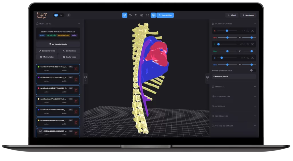
Filum is the comprehensive software solution engineered to bring the precision of medical imaging and 3D modeling to veterinary practice. Designed to address the unique anatomical challenges of each species, the platform transforms CT and MRI scans into high-fidelity, interactive 3D models.
- Oncology treatment simulation
- Pressure and velocity calculation
- Behavior prediction
- Anatomical fidelity at 10-micron resolution
Driving veterinary science
We collaborate with leading institutions on clinical research projects to advance industry knowledge, constantly integrating emerging technologies from our R&D team to elevate the standard of care and innovation in animal health.
Digital twins and high-fidelity biomodels
Digital surgical planning and high-fidelity materialization
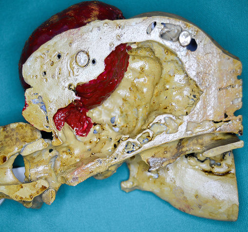
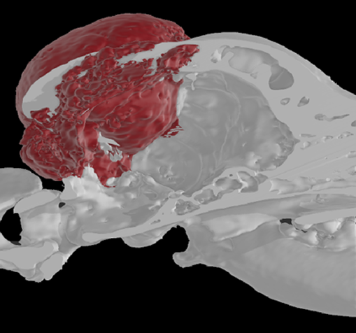
With Filum we transform medical imaging studies into interactive digital twins and high-fidelity physical anatomical replicas. A key technology for planning with absolute accuracy and adapting each procedure to the unique reality of every animal.
Veterinary planning software
- Real-time 1:1 comparison
- Integrated 2D/3D DICOM viewer
- Resection margin simulation
- Dynamic cross-sectioning tools
- Biomechanics calculation and measurements
- Layer-by-layer tissue segmentation
- Annotation and marker management
- Export to physical biomodels
Interactive digital twins
Access exact virtual representations of the animal’s anatomy to analyze margins and practice complex surgeries before entering the operating room.
Custom biomodels and guides
Easily request the physical production of organoids and anatomical models using advanced bioprinting techniques tailored to the anatomy of each species.
Seamless integration
Optimize your processes by reducing traditional planning times from days to just a few hours, without disrupting the internal workflow of your clinic or hospital.
Ready to lead the next generation of surgery?
Optimize your processes, reduce risks, and offer personalized biomechanical engineering solutions.
Frequently Asked Questions
Our technology is multi-species. We regularly work with small and large animals, adapting each design to the patient’s specific bone density and anatomy.
We only require the DICOM files obtained through CT scans or MRI. From these images, our team processes the 3D reconstruction for the analysis and design phase.
By allowing a real visualization of the pathology and the use of custom cutting or drilling guides, the surgeon arrives at the intervention with a predefined plan, minimizing unforeseen events and optimizing the precision of the approaches.
Yes. Our technology allows for differential modeling. We can isolate tumor lesions, vascular malformations, or fractures, assigning them unique visual properties (colors, opacities, or textures) so that the clinical team can study the spatial relationship of the pathology with surrounding critical structures.